
Encephalitis, an inflammation of the brain parenchyma, presents as diffuse and/or focal neuropsychological dysfunction. Although it primarily involves the brain, the meninges are frequently involved (meningoencephalitis).
The cause of encephalitis is usually infectious in nature. Viral agents, such as Herpes simplex encephalitis (HSE) types 1 and 2 (the latter much more common in neonates than adults), Varicella-zoster virus encephalitis (VZVE), Epstein-Barr virus (EBV), measles virus (PIE and SSPE), mumps virus, and rubella virus, are spread through person-to-person contact. Human herpesvirus 6 may also be a causative agent.
From an epidemiologic and pathophysiologic perspective, encephalitis is distinct from meningitis, though on clinical evaluation both can be present, with signs and symptoms of meningeal inflammation, such as photophobia, headache, or stiff neck. It is also distinct from cerebritis. Cerebritis describes the stage preceding abscess formation and implies a highly destructive bacterial infection of brain tissue, whereas acute encephalitis is most commonly a viral infection with parenchymal damage varying from mild to profound. Although bacterial, fungal, and autoimmune disorders can also produce encephalitis.
Bacterial pathogens, such as Mycoplasma species and those causing rickettsial disease or cat scratch disease, are rare and invariably involve inflammation of the meninges out of proportion to their encephalitic components. Encephalitis due to parasites and fungi other than Toxoplasma gondii are covered elsewhere.
Noninfectious causes include the demyelinating process in acute disseminated encephalitis.
Etiology
The incidence of encephalitis with herpes simplex virus (HSV) being the most common cause. Considering the subacute and chronic encephalopathies, the emergency department (ED) physician is most likely to encounter toxoplasmosis in an immune-compromised host.
The relatively common acute arboviral encephalitis vary widely in epidemiology, mortality, morbidity, and clinical presentation. However, attempts to distinguish these acute arboviral encephalitis from the treatable acute viral encephalitis due to herpes simplex or varicella are important.
Herpes simplex encephalitis (HSE), which occurs sporadically in healthy and immune-compromised adults is also encountered in neonates infected at birth during vaginal delivery and is potentially lethal if not treated. Varicella-zoster virus encephalitis (VZVE) is life threatening in immune-compromised patients. Swift identification and immediate treatment of HSE or VZVE can be lifesaving. From a risk-benefit standpoint, most authorities recommend initiating emergency department allopathic treatment with acyclovir in any patient whose central nervous system (CNS) presentation is suggestive of viral encephalitis, especially in the presence of fever, encephalopathy, or focal findings, and in all neonates who appear ill for whom a central nervous system (CNS) infection is being considered.
See the following for more clinical information: 
- California Encephalitis.
- CBRNE–Venezuelan Equine Encephalitis.
- Eastern Equine Encephalitis.
- Herpes Simplex Encephalitis.
- HIV-Associated Cytomegalovirus Encephalitis.
- Japanese Encephalitis.
- St. Louis Encephalitis.
- Venezuelan Equine Encephalitis.
- Viral Encephalitis.
- West Nile Encephalitis.
- Western Equine Encephalitis.
Pathophysiology
Portals of entry are virus specific. Many viruses are transmitted by humans, though most cases of Herpes simplex encephalitis (HSE) are thought to be reactivation of HSV lying dormant in the trigeminal ganglia. Mosquitoes or ticks inoculate arbovirus, and rabies virus is transferred via an infected animal bite or exposure to animal secretions. With some viruses, such as varicella-zoster virus (VZV) and cytomegalovirus (CMV), an immune-compromised state is usually necessary to develop clinically apparent encephalitis.
In general, the virus replicates outside the CNS and gains entry to the CNS either by hematogenous spread or by travel along neural pathways (eg, rabies virus, HSV, VZV). The etiology of slow virus infections, such as those implicated in the measles-related subacute sclerosing panencephalitis (SSPE) and progressive multifocal leukoencephalopathy (PML), is poorly understood.
Once across the blood-brain barrier, the virus enters neural cells, with resultant disruption in cell functioning, perivascular congestion, hemorrhage, and a diffuse inflammatory response that disproportionately affects gray matter over white matter. Regional tropism associated with certain viruses is due to neuron cell membrane receptors found only in specific portions of the brain, with more intense focal pathology in these areas. A classic example is the herpes simplex virus predilection for the inferior and medial temporal lobes.
In contrast to viruses that invade gray matter directly, acute disseminated encephalitis and postinfectious encephalomyelitis (PIE), most commonly due to measles infection and associated with Epstein-Barr virus (EBV) and cytomegalovirus (CMV) infections, are immune-mediated processes that result in multifocal demyelination of perivenous white matter.
Epidemiology of Encephalitis
Determining the true incidence of encephalitis is impossible, because reporting policies are neither standardized nor rigorously enforced.
Herpes simplex encephalopathy (HSE), the most common cause of sporadic encephalitis in Western countries, is relatively rare; the overall incidence is 0.2 per 100,000, with neonatal HSV infection occurring in 2-3 per 10,000 live births.
The arbovirus group is the most common cause of episodic encephalitis, with a reported incidence similar to that of Herpes simplex virus (HSV). These statistics may be misleading in that most people bitten by arbovirus-infected insects do not develop clinically apparent illness and, of those who do, less than 10% develop overt encephalitis. Arboviruses require an insect vector.
Among the other arbovirus-caused encephalitis, the deadliest (and, fortunately, rarest) is eastern equine encephalitis (EEE), which is encountered in New England and surrounding areas.
Western equine encephalitis (WEE), a milder disease, is most common in rural communities.
Powassan virus is the only well-documented arbovirus transmitted by ticks.
Less common causes of viral encephalitis include VZV encephalitis, with an incidence of roughly 1 in 2000 infected persons.
Measles produces 2 devastating forms of encephalitis: PIE, which occurs in about 1 in 1000 infected persons, and SSPE, occurring in about 1 in 100,000 infected patients.
Rarest cases are of rabies encephalitis, typically a consequence of the immigration of an infected bird or an animal to the area.
International statistics
Japanese virus encephalitis (JE), occurring principally in Japan, Iran, Iraq, Bangladesh, Nepal, Afghanistan, Pakistan, Taiwan, Indonesia, Phippen, Vietnam, Thailand, Myanmar, Cambodia, East Taimor, Brunei, Laos, Singapore, Malaysia, China and India, is the most common viral encephalitis.
Age-related differences in incidence
Individuals at the extremes of age are at highest risk, particularly for HSE. Neonatal HSE is a 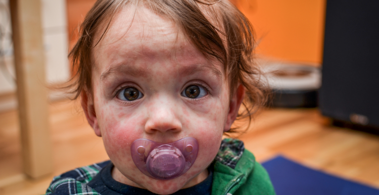 manifestation of disseminated infection type 1 or 2, whereas older infants, children, and adults are much more likely to have localizing CNS infection almost exclusively due to type 1, in a bimodal distribution of patients aged 5-30 years or older than 50 years.
St Louis encephalitis and west Nile encephalopathy (WNE) are more common and are most severe in patients older than 60 years; conversely.
La Crosse encephalitis virus (LACV) is more common and is most severe in children younger than 16 years.
Eastern equine encephalitis (EEE) and Western equine encephalitis (WEE) disproportionately affect infants while EEE disproportionately affects children and elderly persons.
Signs and symptoms of Encephalitis
The viral prodrome typically consists of fever, headache, nausea and vomiting, lethargy, and myalgias. Manifestations associated with specific types of encephalitis include the following:
- Encephalitis caused by varicella-zoster virus (VZV), Epstein-Barr virus (EBV), cytomegalovirus (CMV), measles virus, or mumps virus: Rash, lymphadenopathy, hepatosplenomegaly, and parotid enlargement,
- St Louis encephalitis: Dysuria and pyuria,
- West Nile encephalitis (WNE): Extreme lethargy
The classic presentation is encephalopathy with diffuse or focal neurologic symptoms, including the following:
- Behavioral and personality changes, with decreased level of consciousness,
- Neck pain, stiffness,
- Photophobia,
- Lethargy,
- Generalized or focal seizures (60% of children with California virus encephalitis [CE]),
- Acute confusion or amnestic states,
- Flaccid paralysis (10% of patients with WNE).
The signs of encephalitis may be diffuse or focal. Typical findings include the following:
- Altered mental status,
- Personality changes (very common),
- Focal findings (eg, hemiparesis, focal seizures, and autonomic dysfunction),
- Movement disorders (eg, St Louis encephalitis, eastern equine encephalitis, and western equine encephalitis),
- Ataxia,
- Cranial nerve defects,
- Dysphagia, particularly in rabies,
- Meningismus (less common and less pronounced than in meningitis),
- Unilateral sensorimotor dysfunction (postinfectious encephalomyelitis).
Findings of herpes simplex virus (HSV) infection in neonates may include the following:
- Herpetic skin lesions over the presenting surface from birth or with breaks in the skin, such as
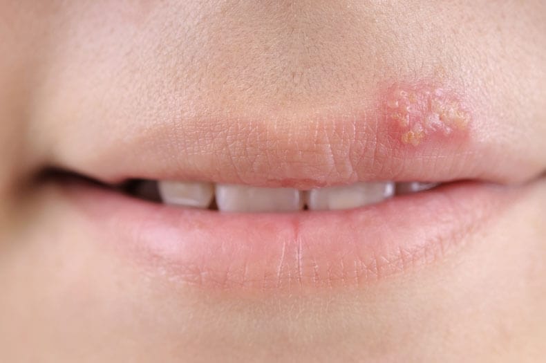 those resulting from fetal scalp monitors,
 - Keratoconjunctivitis,
- Oropharyngeal involvement, particularly buccal mucosa and tongue,
- Encephalitis symptoms (eg, seizures, irritability, change in attentiveness, and bulging fontanelles).
Additional signs of disseminated, severe HSV include jaundice, hepatomegaly, and shock.
Other complains
Encephalitis may be associated with a number of complications, including the following:
- Seizures,
- Syndrome of inappropriate secretion of antidiuretic hormone (SIADH),
- Increased intracranial pressure (ICP),
- Coma.
Diagnosis
Although bacterial, fungal, and autoimmune disorders can produce encephalitis, most cases are viral in origin. Accordingly, in addition to standard blood and urine tests, studies may be performed to identify the infectious agent causing the encephalitis. It is important, when possible, to distinguish acute arboviral encephalitis from potentially treatable acute viral encephalitis, especially herpes simplex encephalitis (HSE) and varicella-zoster encephalitis, as a high suspicion for these disorders and prompt treatment can reduce the severity of neurological sequelae and can be lifesaving.
Physical Examination
Look for supporting evidence of viral infection. The signs of encephalitis may be diffuse or focal. At the  extremes, 80% of patients with HSE present with focal findings. Typical findings include the following:
- Altered mental status,
- Personality changes are very common,
- Focal findings, such as hemiparesis, focal seizures, and autonomic dysfunction,
- Movement disorders (St Louis encephalitis, eastern equine encephalitis [EEE], western equine encephalitis [WEE]),
- Ataxia,
- Cranial nerve defects,
- Dysphagia, particularly in rabies,
- Meningismus (less common and less pronounced than in meningitis),
- Unilateral sensorimotor dysfunction (postinfectious encephalomyelitis [PIE]).
Infants
Findings of HSV infection in neonates (aged 1-45 d) may include the following:
- Herpetic skin lesions over the presenting surface from birth or with breaks in the skin, such as those resulting from fetal scalp monitors,
- Keratoconjunctivitis,
- Oropharyngeal involvement, particularly buccal mucosa and tongue,
- Encephalitis symptoms, such as seizures, irritability, change in level of attentiveness, bulging fontanelles,
- Additional signs of disseminated, severe HSV include jaundice, hepatomegaly, and shock.
As noted above, Toxoplasma infection causing encephalitis is found in immune-suppressed patients. They exhibit significant encephalopathy with lethargy or personality changes, and 75% present may present with focal neuropathology.
Blood and Urine Tests
A complete blood count (CBC) with differential should be performed, although findings are often within the normal range.
Serum electrolyte levels are usually normal unless dehydration is present; the syndrome of inappropriate secretion of antidiuretic hormone (SIADH) occurs in 25% of patients with St Louis encephalitis.
The serum glucose level should be determined to rule out confusion due to treatable hypoglycemia and to compare with the cerebrospinal fluid (CSF) glucose value. Low serum results are found in nutritionally deprived patients, while diabetic patients may present with elevated glucose levels compatible with complicating hyperosmolar state or diabetic ketoacidosis.
Blood urea nitrogen (BUN) and creatinine levels are helpful to assess hydration status, and liver function tests should be performed to assess for end-organ dysfunction or the need to adjust antimicrobial therapy dosing regimens.
A lumbar puncture (LP) should be performed on all patients suspected of having a viral encephalitis. A platelet count and coagulation profile are indicated in patients who are alcohol users, have liver disease, and those in whom disseminated intravascular coagulation (DIC) is suspected. The patient may require platelets or fresh frozen plasma (FFP) before LP.
Urine
A urinary electrolyte test should be performed if SIADH is suspected. Urine or serum toxicology screening may be indicated in selected patients presenting with a toxic delirium or confusional state.
Herpes simplex virus (HSV) cultures of suspicious lesions and a Tzanck smear should be obtained. Viral cultures of CSF, including HSV, should be performed, although the incidence of the latter being positive is rare. Blood cultures for bacterial pathogens should be obtained.
Complement fixation antibodies are useful in identifying arbovirus. Cross-reactivity exists among a subgroup of arboviruses, the flaviviruses (eg, viruses that cause St Louis encephalitis, Japanese virus encephalitis (JE), and West Nile encephalitis (WNE), and the antibodies found in persons inoculated with yellow fever vaccine.
Heterophile antibody and cold agglutinin testing for Epstein-Barr virus (EBV) may be helpful.
Serologic tests for toxoplasmosis can be helpful in light of an abnormal computed tomography (CT) scan, particularly in the case of single lesions. However, the overlap in titer levels between exposed but currently uninfected and reactivated groups may complicate interpretation.
Computed Tomography, Magnetic Resonance Imaging, and Electroencephalography
Performance of a head CT scan with and without contrast agent should be performed in virtually all patients with encephalitis. This should be done prior to LP if there are focal complaints or findings, signs to search for evidence of elevated intracranial pressure (ICP), obstructive hydrocephalus, or mass effect due to focal brain infection. Head CT scanning also helps exclude brain hemorrhage or infarction as a cause of an encephalopathic state. Magnetic resonance imaging (MRI) is more sensitive than CT scanning in demonstrating brain abnormalities earlier in the disease course.
In HSE, MRI may show several foci of increased T2 signal intensity in medial temporal lobes and inferior frontal gray matter. Head CT commonly shows areas of edema or petechial hemorrhage in the same areas. EEE and tick-borne encephalitis may show similar increased MRI signal intensity in the basal ganglia and thalamus.

In toxoplasmosis, contrast-enhanced head CT typically reveals several nodular or ring-enhancing lesions. Because lesions may be missed without contrast, MRI should be performed in patients for whom use of contrast material is contraindicated.
In HSE, electroencephalography (EEG) often documents characteristic paroxysmal lateral epileptiform discharges (PLEDs), even before neuroradiography changes. Eventually, PLEDs are positive in 80% of cases; however, the presence of PLEDs is not pathognomonic for HSE.
Cerebro spinal fluid
A lumbar puncture (LP) should be performed in all cases of suspected viral encephalitis. Studies that may be ordered to identify the infectious agent include the following:
- HSV cultures of suspicious lesions and a Tzanck smear,
- Viral cultures of CSF, including HSV,
- Blood cultures for bacterial pathogens,
- Complement fixation antibodies to identify arbovirus,
- Heterophile antibody and cold agglutinin testing for EBV,
- Serologic tests for Toxoplasma.
Typical patterns of findings in the CSF pressure and CSF analysis follow in the table regarding bacterial versus viral versus fungal (including cryptococcal) meningitis or encephalitis.
Table. Cerebrospinal Fluid Findings by Type of Organism
| CSF Finding (Normal) | Bacterial Meningitis | Viral Meningitis* | Fungal Meningitis |
| Pressure (5-15 cm water) |
|
|
|
| Cell counts, mononuclear cells/µL
Preterm (0-25) Term (0-22) 6 mo+ (0-5) |
|
|
|
| Microorganisms (none) |
|
|
|
| Glucose†
Euglycemia (>50% serum) Hyperglycemia (>30% serum) |
|
|
|
| Protein
Preterm (65-150 mg/dL) Term (20-170 mg/dL 6 mo+ (15-45 mg/dL) |
|
|
|
| *Some bacteria (eg, Mycoplasma, Listeria, Leptospira, Borrelia burgdorferi [Lyme disease]) cause alterations in spinal fluid that resemble the viral profile. An aseptic profile is also typical of partially treated bacterial infections (>33%, especially those in children, are treated with antimicrobials) and of the 2 most common causes of encephalitis—the arboviruses and the potentially curable HSV.
† Wait 4 hours after glucose load. AFB—acid-fast bacillus; CSF—cerebrospinal fluid; EEE-eastern equine encephalitis; HSV—herpes simplex virus; RBC—red blood cell; TB—tuberculosis; WBC—white blood cell. |
|||
Gram staining
The most important diagnostic test in the emergency department (ED) to rule out bacterial meningitis is prompt Gram staining and, if available, polymerase chain reaction (PCR) of the CSF in patients with suspected HSV encephalitis. PCR for HSV DNA is 100% specific and 75-98% sensitive within the first 25-45 hours. Types 1 and 2 cross-react, but no cross-reactivity with other herpes viruses occurs. Arguably, a series of quantitative PCRs documenting the decline of viral load with acyclovir treatment is strongly supportive of the diagnosis of HSV, and selected patients my avoid need for brain biopsy.
Allopathic treatment for encephalitis
No satisfactory allopathic treatment exists for encephalitis infections. The goals of  pharmacotherapy are to reduce morbidity and prevent complications. Antivirals are used to manage treatable viral encephalitis. Corticosteroids may be considered for postinfectious or noninfectious encephalitis.
In the prehospital setting, evaluate and treat for shock or hypotension. Administer crystalloid infusion in patients with evidence of circulatory compromise. Consider airway protection in patients with an altered mental status. Seizure precautions are indicated. Treat seizures according to usual protocols (ie, lorazepam 0.1 mg/kg given intravenously [IV]). All patients should receive oxygen and have intravenous access secured en route to the emergency department (ED).
In the emergency department (ED), beyond supportive care, viral encephalitis is not treatable, with the exceptions of HSV and VZV encephalitis. Important initial measures include the following:
-
Administration of the first dose or doses of acyclovir, with or without antibiotics or steroids, as quickly as possible; the standard for acute bacterial meningitis is initiation of treatment within 30 minutes of arrival
-
Consideration of an ED triage protocol to identify patients at risk for HSV encephalitis
-
Collection of laboratory samples and blood cultures before the start of IV therapy
-
Neuroimaging (eg, MRI or, if that is unavailable, contrast-enhanced head CT) before LP
Management of hydrocephalus and increased intracranial pressure
In patients with hydrocephalus and increased intracranial pressure (ICP), general measures include management of fever and pain, control of straining and coughing, and prevention of seizures and systemic hypotension.
In otherwise stable patients, elevating the head and monitoring neurologic status usually are sufficient. When more aggressive maneuvers are indicated, early use of diuresis (eg, furosemide 20 mg IV, mannitol 1 g/kg IV) may be helpful, provided that circulatory volume is protected. Dexamethasone 10 mg IV q6h helps in managing edema surrounding space-occupying lesions. Hyperventilation (arterial CO2 tension [PaCO2] 30 mm Hg) may cause a disproportional decrease in cerebral blood flow (CBF), but it is used to control increasing ICP on an emergency basis.
Intraventricular ICP monitoring is controversial. Some authorities believe that dangerous focal edema with a pressure gradient between the temporal lobe and the sub tentorial space usually is not detected by the monitor and that this failure of detection can lead to a false sense of security. In fact, monitor placement may potentially aggravate a pressure gradient.
Allopathic treatment of systemic complications of Encephalitis
Look for and treat systemic complications (eg, hypotension or shock, hypoxemia, hyponatremia, and exacerbation of chronic diseases), particularly in herpes simplex encephalitis (HSE), eastern equine encephalitis (EEE), Japanese virus encephalitis (JE).
Empiric allopathic treatment of HSV meningoencephalitis and VZV encephalitis
Empiric adult emergency treatment for herpes simplex virus (HSV) meningoencephalitis and varicella-zoster virus (VZV) encephalitis consists of acyclovir 10 mg/kg (infused over 1 h) q8h for 14-21 days. Give acyclovir 10-15 mg/kg IV q8h for neonatal HSV; for HSV encephalitis in the pediatric population, give acyclovir 10 mg/kg IV q8h.
In HIV-positive patients, consider foscarnet, given the increased incidence of acyclovir-resistant HSV and herpes zoster virus (HZV).
Complications pre allopathic treatment of Encephalitis
Encephalitis may be associated with a number of complications, including the following:
- Seizures,
- Syndrome of inappropriate secretion of antidiuretic hormone (SIADH),
- Increased intracranial pressure (ICP),
- Coma.
Homeopathic treatment for Encephalitis
In Homeopathic medicines there are many best and proven medicines for many viral infections, here 
The symptoms listed against each medicine may not be directly related to this disease because in homeopathy general symptoms and constitutional indications are taken into account for selecting a remedy.
Hydrophobinum
Hydrophobinum – the nosode of rabies. Bubo. Clairvoyance. Convulsions. Sparks before eyes and vanishing of sight; red face; involuntary grinding of teeth. Corns, and its pains. Diarrhea. Dysentery. Fever. Hair oiliness. Headache. Hydrophobia. Hypersensitiveness. Landry’s paralysis. Lyssophobia. Mania. Nervousness. Neuralgia. Esophageal stricture. Paralysis. Convulsions. Respiratory paralysis. Salivation. Satyriasis. Sciatica. Sunstroke. Tetanus. Ulcers. Vaginismus.
Lyssinum
Affects principally the central and peripheral nervous system. Aching in bones. Abnormal sexual desire. Convulsions brought on by dazzling light or sight of running water. Lyssophobia; fear of becoming mad. Emotion and bad news aggravate; also, thinking of fluids. Hypersensitiveness of all senses. Chronic headache. Boring pain in forehead. Constant spitting; saliva tough, viscid. Sore throat: constant desire to swallow, which is difficult; gagging when swallowing water. Froths at mouth.
Cantharis Vesicatoria
A powerful medicine. Violent inflammations. Frenzied delirium. Furious delirium. Anxious restlessness, ending in rage. Hydrophobia. Puerperal convulsions. Violent inflammation of the whole gastro-intestinal canal, especially lower bowel. Over sensitiveness of all parts. Irritation. Raw, burning pains. Hemorrhages. Intolerable, constant urging to urinate. Dysuria. Increases secretion of mucous membranes, tenacious mucus. The inflammations cantharis produces (bladder, kidneys, ovaries, meninges, pleuritic and pericardial membranes).
Constantly attempts to do something but accomplishes nothing. Acute mania, generally of a sexual type; amorous frenzy; fiery sexual desire. Paroxysms of rage, crying, barking. Sudden loss of consciousness with red face. Burning like boiling in brain. Vertigo.
Acid Nitricum
Irritable, hateful, vindictive, headstrong. Hopeless despair. Sensitive to noise, pain, touch, jar. Fear of death. Sensation of a band around head. Headache from little pressure; full feeling; worse from noises. Scalp sensitive. Difficult hearing. Very sensitive to noise. Cracking in ears when chewing. Double vision; sharp, sticking pains. Photophobia, constant lachrymation.
Fagus sylvatica
Epilepsy. Headache. Hydrophobia. Vertigo. Dread of liquids and salivation. Nerve irritation. 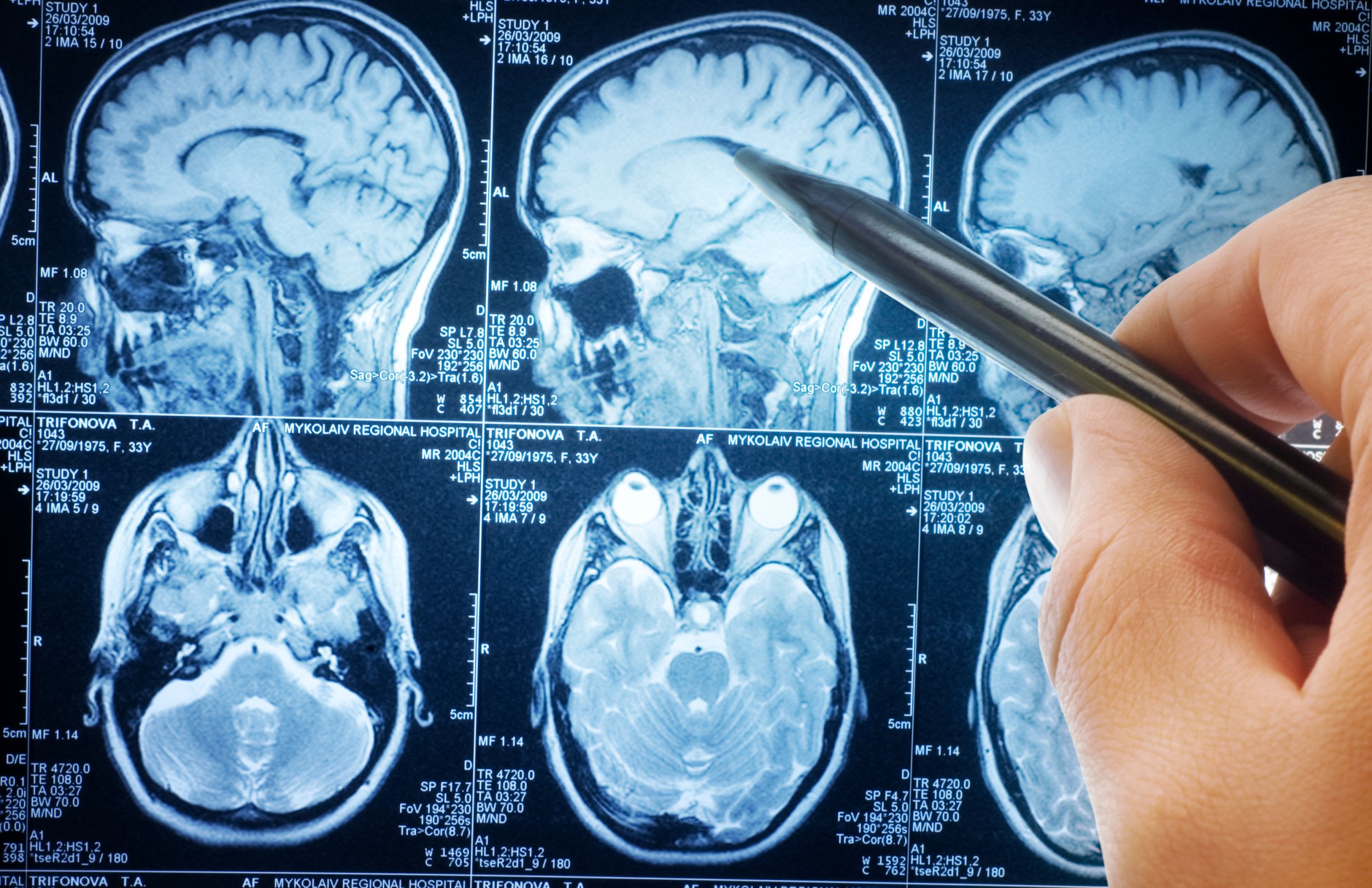 Hydrophobia, trembling, convulsions with periodic spasms, stiffness and coldness. Swelling of the mouth. Coma due to Cerebro spinal infections. Froth and saliva; intolerable thirst; entreating for drink, but as soon as any liquid was brought, he seemed to shudder as after eating unripe grapes. Seized with gloominess, torpor, and dread of liquids; urine fiery red, depositing copious turbid white sediment. A few hours before death vomited a voracious bile. Nausea. Dizziness and stupefaction. Swelling of mouth.
Coffea Cruda
Great nervous agitation and restlessness. Extreme sensitiveness characterizes this remedy. Neuralgia in various parts; always with great nervous excitability and intolerance of pain, driving to despair. Unusual activity of mind and body. Bad effects of sudden emotions, surprises, joy, etc. Nervous palpitation. Coffea is specially suited to tall, lean, stooping persons with dark complexions, temperament choleric and sanguine. Skin hypersensitive.
Mind: Gaiety, easy comprehension, irritability, excited; senses acute. Impressionable, especially to pleasurable impressions. Full of ideas, quick to act. Tossing about in anguish (Acon).
Head: Tight pain, worse from noise, smell, narcotics. Seems as if brain were torn to pieces, as if nail were driven in head. Worse in open air. Sensitive hearing.
Agave Americana (American Aloe or Century plant)
Gonorrhea. Hydrophobia. Scurvy. Stomachache. Quarrelsomeness, excitement, fright, inability to swallow, small frequent pulse, great anxiety. Biting other people. Violent nervous attacks. Burning in mouth. Painful erections in gonorrhea.
Nux Vomica
Very irritable. Over sensitiveness – sensitive to all impressions. Ugly, malicious. Cannot bear noises, odors, light, etc. Does not want to be touched. Sullen, fault-finding. Headache in occiput or over eyes, with vertigo; brain feels turning in a circle. Vertigo, with momentary loss of consciousness. Intoxicated feeling. Pressing pain on vertex – like nail driven in. Scalp sensitive. Frontal headache, with desire to press the head against something. Congestive headache, associated with hemorrhoids.
Photophobia. Smarting dry sensation in inner canthi. Infra-orbital neuralgia, with watering of eyes. Optic nerve atrophy, from intoxicants. Paresis of ocular muscles. Orbital twitching radiating towards the occiput, Optic neuritis.
Itching in ear through Eustachian tube. Auditory canal dry and sensitive. Otalgia – worse in bed. Hyperesthesia of auditory nerves; loud sounds are painful and anger him.
Lycopodium Clavatum
Melancholy; afraid to be alone. Little things annoy, extremely sensitive. Averse to undertaking new 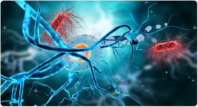 things. Head strong and haughty when sick. Loss of self-confidence. Hurried when eating. Constant fear of breaking down under stress. Apprehensive. Weak memory confused thoughts; spells or writes wrong. Cannot bear to see anything new. Cannot read what he writes. Sadness.
Shakes head without apparent cause. Twists face and mouth. Pressing headache on vertex; worse from lying down or stooping. Throbbing headache after every paroxysm of coughing. Headaches over eyes in severe colds; better, uncovering. Vertigo. Pain in temples. Eczema; moist oozing behind ears. Deep furrows on forehead. Blindness, night-blindness more characteristic. Sees only one-half of an object.
Thick, yellow, offensive discharge from ears. Otorrhea and deafness with or without tinnitus; after scarlatina. Humming and roaring with hardness of hearing; every noise causes peculiar echo in ear.
Anacardium Orientale
Neurasthenics; impaired memory, depression, and irritability; diminution of senses (smell, sight, hearing). Syphilitic patients. Sensation of a plug in various parts-eyes, rectum, bladder, etc. Fixed ideas. Hallucinations: thinks he is possessed of two persons or wills. Anxiety when walking. Profound melancholy and hypochondriasis, with tendency to use violent language. Brain-fag. Impaired memory. Absent mindedness. Very easily offended. Malicious; seems bent on wickedness. Lack of confidence in himself or others. Suspicious. Clairaudient hears voices far away or of the dead. Senile dementia. Absence of all moral restraint.
Vertigo. Pressing pain, as from a plug. Itching and little boils on scalp. Pressure like a plug on upper orbit. Indistinct vision. Objects appear too far off. Pressing in the ears as from a plug. Hard of hearing.
Belladonna
Belladonna acts upon every part of the nervous system. Congestion, furious excitement, perverted special senses, twitching, convulsions and pain. Throbbing carotids, excited mental state, hyperesthesia of all senses, delirium, restless sleep, convulsive movements, dryness of mouth and throat with aversion to water, neuralgic pains that come and go suddenly. Heat, redness, throbbing and burning. Epileptic spasms.
Scarlet fever and also prophylactic. Exophthalmic goiter – extreme of thyroid toxemia. No thirst. Anxiety and fear. Violence of attack and suddenness of onset. Patient lives in a world of his own, engrossed by specters and visions and oblivious to surrounding realities. Retina insensible to actual objects, a host of visual hallucinations throng about him and come to him from within. Visual impressions and fantastic illusions. Hallucinations; sees monsters, hideous faces. Delirium; frightful images; furious; rages, bites, strikes; desire to escape. Loss of consciousness. Disinclined to talk. Perversity, with tears. Acuteness of all senses. Changeableness.
Vertigo, with falling to left side or backwards. Sensitive to least contact. Throbbing and heat. Palpitation reverberating in head with labored breathing. Boring of head into pillow; drawn backward and rolls from side to side. Constant moaning. Face red, bluish-red, hot, swollen, shining; convulsive motion of muscles of face. Swelling of upper lip. Facial neuralgia with twitching muscles and flushed face.
Throbbing deep in eyes on lying down. Pupils dilated. Eyes swollen and protruding, staring, brilliant; conjunctiva red; dry, burn; photophobia; shooting in eyes. Exophthalmos. Ocular illusions; fiery appearance. Diplopia, squinting, spasms of lids. Fundus congested.
Tearing pain in middle and external ear. Humming noises. Membrana tympani bulges and injected. Parotid gland swollen. Sensitive to loud tones. Hearing very acute. Otitis media. Pain causes delirium. Hematoma auris. Acute and sub-acute conditions of Eustachian tube. Autophony-hearing one’s voice in ear.
Medorrhinum
Great disturbance and irritability of nervous system. Pains intolerable; tensive; nerves quiver and  tingle. Dwarfed and stunted. Chronic catarrhal conditions. State of collapse and trembling all over. History of sycosis. Intensity of all sensations. Weak memory. Loses the thread of conversation. Cannot speak without weeping. Time passes too slowly. Difficult concentration. Fears going insane. Sensibility exalted. Nervous, restless. Fear in the dark and of some one behind. Melancholy, with suicidal thoughts.
Burning pain in brain; worse, occiput. Head heavy and drawn backward. Headache from jarring of cars, exhaustion, or hard work. Weight and pressure in vertex. Itching of scalp. Feels as if she stared at everything. Eyeballs ache. Feels as if sticks in eyes. Lids irritated. Partial deafness, pulsation in ears. Quick, darting pains in right ear.
Mercuris Corisive
Delirium, stupor. Frontal pain, congestion of head with burning in cheeks. Drawing pain in periosteum of skull. Pain behind eyeballs. Phlyctenulae; deep ulcers on cornea. Excessive photophobia and acrid lachrymation. Iritis, ordinary or syphilitic. Pain severe at night; burning, shooting, tearing. Little tendency to pus formation. Iris muddy in color, thick, and neither contracts nor dilates. Retinitis albuminuria, ophthalmia neonatorum. Lids edematous, red, excoriated. Severe burning. Soreness of the eyes.
Stramonium
The entire force of this medicine seems to be expended on the brain. Suppressed secretions and excretions. Sensation as if limbs were separated from body. Delirium tremens. Absence of pain and muscular mobility especially of muscles of expression and of locomotion. Gyratory and graceful motions. Parkinson disease.
Devout, earnest, beseeching and ceaseless talking. Loquacious, garrulous, laughing, singing, swearing, praying, rhyming. Sees ghosts, hears voices, talks with spirits. Rapid changes from joy to sadness. Violent and lewd. Delusions about his identity; thinks himself tall, double, a part missing. Religious mania. Cannot bear solitude or darkness; must have light and company. Sight of water or anything glittering brings on spasms. Delirium, with desire to escape.
Raises head frequently from the pillow. Pain in forehead and over eyebrows. Boring pain, preceded by obscure vision. Rush of blood to head; staggers, with tendency to fall forward and to the left. Auditory hallucinations. Seem prominent, staring wide open; pupils dilated. Loss of vision; complains that it is dark and calls for light. Small objects look large. Parts of the body seem enormously swollen. Strabismus. All objects look black.
Arsenicum Album
Anguish and restlessness, changes place continually. Fears, of death, of being left alone, fear with cold sweat. Suicidal. Hallucinations of smell and sight. Despair drives him from place to place. Miserly, malicious, selfish, lacks courage. General sensibility increased. Sensitive to disorder and confusion.
Headaches, periodical burning pains, with restlessness; with cold skin. Hemicrania, with icy feeling of scalp and great weakness. Sensitive head in open air. Delirium tremens; cursing and raving; vicious. Head is in constant motion. Scalp itches intolerably; circular patches of bare spots; rough, dirty, sensitive, and covered with dry scales; nightly burning and itching; dandruff. Scalp very sensitive; cannot brush hair.
Burning in eyes. Acrid lachrymation. Lids red, ulcerated, scabby, scaly, granulated. External inflammation, with extreme painfulness; burning, hot, and excoriating lachrymation. Corneal ulceration. Intense photophobia. Ciliary neuralgia, with fine burning pain.
Ears – Skin within, raw and burning. Thin, excoriating, offensive otorrhea. Roaring in ears, during a paroxysm of pain.
Chamomilla
Anger. Asthma from anger. Blepharospasm. Convulsions. Waking, screaming on. Peevishness, restlessness. Blepharitis; ophthalmia. Screaming. Sensitiveness irritable, thirsty, hot, and numb. Erysipelas. Excitement. Excoriation. Fainting fits. Fevers. Mumps. Neuralgia. Parotitis. Perichondritis. Peritonitis. Tinnitus. Speech, affections. Lientery. Labour: disorders of; after-pains. Mastitis. Salivation. Spasms. Impatient, intolerant of being spoken to or interrupted; extremely sensitive to every pain; always complaining. Throbbing headache.
Lachesis Mutus
Purpura, septic states, diphtheria, and other low forms of disease, when the system is thoroughly poisoned, and the prostration is profound. Delirium tremens with much trembling and confusion. Melancholia. Diphtheritic paralysis (Botulinum). Diphtheria carriers. Sensation of tension in various parts. Cannot bear anything tight anywhere.
Great loquacity. Amative, sad, no desire to mix with the others. Restless and uneasy; does not wish to attend to business; wants to be off somewhere all the time. Jealous. Euthanasia. Suspicious; nightly delusion of fire. Religious insanity. Derangement of the time sense. 
Headaches – pain at root of nose, pressure and burning on vertex, waves of pain; worse after moving. Sun headaches. With headache, flickering, dim vision, very pale face. Vertigo. Defective vision after diphtheria, extrinsic muscles too weak to maintain focus. Sensation as if eyes were drawn together by cords which were tied in a knot at root of nose. Tearing pain from zygoma into ear; also, with sore throat. Ear-wax hard, dry.
Veratrum Album
A perfect picture of collapse, with extreme coldness, blueness, and weakness. Surgical shock. Infection – shock with cold sweat on forehead, pale face, rapid, feeble pulse. Cold perspiration. Profuse vomiting, purging, and cramps in extremities. Excessive dryness of all mucous surfaces. “Coprophagia” violent mania alternates with silence and refusal to talk.
Melancholy, with stupor and mania. Sits in a stupid manner; notices nothing; Sullen indifference. Frenzy of excitement; shrieks, curses. Puerperal mania. Aimless wandering from home. Delusions of impending misfortunes. Mania, with desire to cut and tear things. Attacks of pain, with delirium driving to madness. Cursing, howling all night.
Contracted features. Cold sweat on forehead. Sensation of a lump of ice on vertex. Headache, with nausea, vomiting, diarrhea, pale face. Neck too weak to hold head up.
Hyoscyamus Niger
Profoundly disturbed nervous system, as if some diabolical force took possession of the brain and prevented its functions. Mania of a quarrelsome and obscene character. Inclined to be unseemly and immodest in acts, gestures and expressions. Very talkative, and persists in stripping herself, or uncovering genitals. Jealous, afraid of being poisoned, etc. Weakness and nervous agitation (typhoid and other infections with coma vigil). Tremulous weakness and twitching of tendons. Subsalts tendendum. Muscular twitching, spasmodic affections, generally with delirium. Delirium, with attempt to run away. Non-inflammatory cerebral activity. Toxic gastritis.
Very suspicious. Great hilarity; inclined to laugh at everything. Low, muttering speech; constant carphologia, deep stupor. Vertigo, brain feels loose, fluctuating. Inflammation of brain, with unconsciousness; head is shaken to and fro. Pupils dilated, sparkling, fixed. Eyes open, but does not pay attention; downcast and dull, fixed. Strabismus. Spasmodic closing of lids. Diplopia. Objects have colored borders.
Prognosis
The prognosis is dependent on the type (viral or bacterial) and stage of infection, treatment type (allopathic or Homeopathic), extremes of age (< 1 y or >55 y), immune-compromised status, and preexisting neurologic conditions.
Allopathically treated encephalitis has a mortality of 50-75%, and virtually all allopathically or untreated or late-treatment survivors have long-term motor and mental disabilities. The mortality in treated encephalopathies averages 20%, and the neurologic outcome correlates with the neurological disability. With allopathy, approximately 40% of survivors have minor-to-major learning disabilities, memory impairment, neuropsychiatric abnormalities, epilepsy, fine-motor-control deficits, and dysarthria.
Results
Outcomes in arboviral JE and EEE are catastrophic, similar to untreated or treated allopathically HSE,  with high mortality and severe morbidity, including intellectual disability, hemiplegia, and seizures. Other arboviruses cause substantially less morbidity and mortality. For example, with allopathic treatment, St Louis encephalitis and WNE have a mortality rate of 2-20%, the higher rates found in patients older than 60 years. Long-term sequelae with St Louis encephalitis include behavioral disorders, memory loss, and seizures.
WEE
WEE is associated with few deaths and much less morbidity; complete health is hundred percent possible with Homeopathic treatment. Developmental delay, seizure disorder, and paralysis occasionally occur in children, and postencephalitic parkinsonism may occur in adults treated allopathically. CE is typically associated with mild illness, and most patients make a full recovery even with allopathy too; however, with allopathy the minority of patients with severe disease have a 25% chance of focal neurologic dysfunction. Death rates from WEE and LAC are less than 5%.
PIE
PIE secondary to measles is associated with a mortality rate (with allopathic treatment) approaching 40% of cases, with a high rate of neurologic sequelae in survivors. SSPE is uniformly fatal, although the disease course may last anywhere from several weeks to 10 years; on other hand with Homeopathic treatment, it is curable completely.
VZVE
VZVE pre allopathic treatment, has a mortality of 15% in immune-competent patients and virtually 100% in immune-suppressed patients, encephalitis mortality rate is 8%, with substantial morbidity found in approximately 12% of survivors. With Homeopathic treatment mortality is <2%.
Rabies encephalitis and acute disseminated encephalitis are virtually 100% fatal pre allopathic treatment, although there are rare survivors reported in the medical literature. While with Homeopathic treatment survivor rate is >68%.
P. S: This article is only for doctors having good knowledge about Homeopathy and allopathy, for learning purpose(s).
For proper consultation and treatment, please visit our clinic.
None of above-mentioned medicine(s) is/are the full/complete treatment but just hints for treatment; every patient has his/her own constitutional medicine.
 Dr. Sayyad Qaisar Ahmed (MD {Ukraine}, DHMS), Abdominal Surgeries, Oncological surgeries, Gastroenterologist, Specialist Homeopathic Medicines.
Senior research officer at Dnepropetrovsk state medical academy Ukraine.
Location: Al-Haytham clinic, Umer Farooq Chowk Risalpur Sadder (0923631023, 03119884588), K.P.K, Pakistan.
Find more about Dr Sayed Qaisar Ahmed at:
https://www.youtube.com/Dr Qaisar Ahmed