
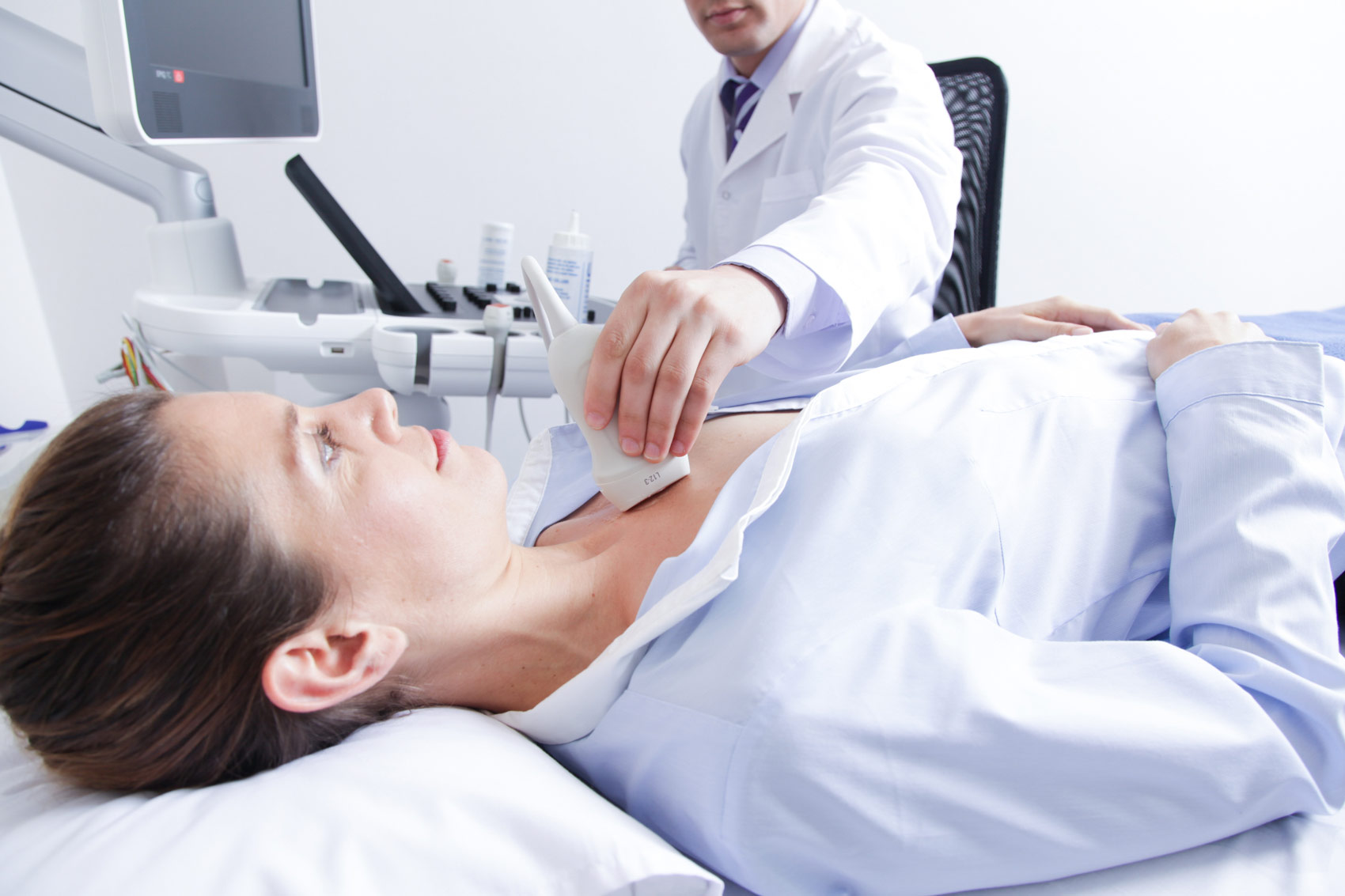
An echocardiogram is an ultrasound test that checks the structure and function of heart. An echo can diagnose a range of conditions including cardiomyopathy and valve disease. There are several types of echo tests, including transthoracic and transesophageal.
What is an echocardiogram?
An echocardiogram (echo) is a graphic outline of our heart’s movement. During an echo test, technician uses ultrasound (high-frequency sound waves) from a hand-held wand placed on patient’s chest to take pictures of his/her heart’s valves and chambers. This helps the provider evaluate the pumping action of heart.
Doctors often combine echo with Doppler ultrasound and color Doppler techniques to evaluate blood flow across patient’s heart’s valves.
Echocardiography uses no radiation. This makes an echo different from other tests like X-rays and CT scans that use huge amounts of radiation.
Who performs an echo test?
A cardiac sonographer (technician) performs echo. They’re trained in performing echo tests using the most current technology. They’re prepared to work in a variety of settings including hospital rooms and catheterization labs.
What are the different types of echocardiograms?
There are several types of echocardiograms. Each one offers unique benefits in diagnosing and managing heart disease. They include:
Transthoracic echocardiogram
 Healthcare providers use TTE in many ways; the tests can:
- Assess heart health before or after diagnosis and treatment.
- Identify the cause of certain symptoms.
- Screen for and diagnose possible medical conditions.
TTE can evaluate heart health by:
- Checking your heart valves.
- Determining how well heart is pumping blood.
- Measuring blood pressure and how quickly blood is flowing through the heart.
- Measuring the size and shape of heart’s chambers.
The test can identify causes of cardiac-related symptoms, such as:
- Chest pain.
- Edema (swelling).
- Heart murmur.
- Shortness of breath (dyspnea).
TTE is useful to screen for, diagnose or follow up on specific medical conditions, including:
- Aortic aneurysm or aortic dissection.
- Blood clots.
- Changes in electrocardiogram (ECG) results.
- Congenital heart conditions.
- Heart failure.
- Heart valve disease.
- Hypertrophic cardiomyopathy.
- Tumors (heart cancer).
Transesophageal echocardiogram
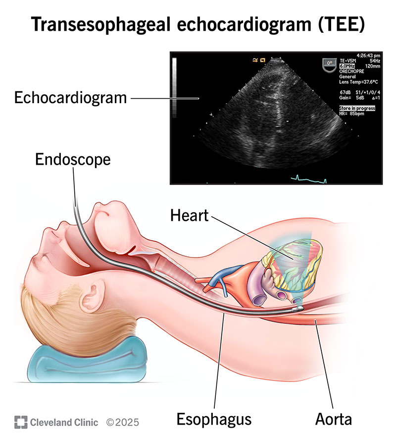
You may need a transesophageal echocardiogram if:
Other tests didn’t show enough detail:
You might’ve had a transthoracic echo, but it wasn’t enough for your provider to diagnose or manage a heart problem. A TEE provides a detailed view of your heart that some other tests can’t.
You’re in critical condition:
A TEE can help your provider learn more if you have very low blood pressure, low oxygen or your body is in shock. It can also help if you have sudden problems with your aorta, like a tear, chest trauma or issues with a prosthetic heart valve.
You’re having a procedure to treat arrhythmia:
Your provider may use TEE before you have catheter ablation or electrical cardioversion. The test can show if you have blood clots in your heart. The clots could break free during the procedure and cause serious problems.
You’re having heart surgery:
TEE is a valuable tool during certain surgeries and procedures. Your provider may use TEE before surgery to check your anatomy. They may use it after surgery to confirm success. TEE can also guide some procedures in real time.
Providers typically do a transthoracic echo first because it’s less invasive. But they may skip that and go right to using a transesophageal echo if you have:
- Chest anatomy that makes a transthoracic echo difficult,
- Lung disorders,
- A life-threatening emergency.
Exercise stress echocardiogram
An exercise stress echocardiogram is an ultrasound of your heart taken before and after exercise. This test is also commonly called stress echo or echo stress test. It produces moving images that show how well your heart functions under stress. Providers use this test to diagnose or monitor heart conditions and ensure you get the treatment you need.
An echocardiogram, or echo, is a test that shows how well your heart is working. It uses sound waves (ultrasound) to produce moving images of your heart. These images allow your provider to see the shape, size and motion of your heart and valves.
An exercise stress echo assesses your heart’s function when it’s beating fast. You create this “stress” by exercising on a treadmill or bicycle.
Are there other types of stress tests?
 Other types of exercise stress tests include:
Exercise stress electrocardiogram:
This test uses an electrocardiogram (EKG) to measure the electrical activity of your heart while you exercise.
Nuclear exercise stress test:
In this test, you receive a small injection of a radioactive substance into a blood vessel. Special imaging before and after exercise detects the radiation and highlights blood flow to your heart.
For people who can’t exercise, there are two alternate ways to create stress on the heart. Providers use these methods for stress echocardiograms, EKGs and nuclear tests:
Pharmacological stress test:
This test uses medications, including dobutamine and vasodilators, to produce stress. Dobutamine increases the heart rate. Vasodilators expand the blood vessels, which increases blood flow through the heart.
Pacing stress test:
You may receive this test if you have a permanent pacemaker to help stabilize your heart rhythms. Your provider programs the device to raise your heart rate during the stress test.
What techniques are used in echocardiography?
Several techniques can be used to create pictures of heart. The best technique depends on patient’s specific condition and what a doctor needs to see. These techniques include:
- Two-dimensional (2D) ultrasound. This approach is used most often. It produces 2D images that appear as “slices” on the computer screen. Traditionally, these slices could be “stacked” to build a 3D structure.
- Three-dimensional (3D) ultrasound. Advances in technology have made 3D imaging more efficient and useful. New 3D techniques show different aspects of heart, including how well it pumps blood, with greater accuracy. Using 3D also allows sonographer to see parts of the heart from different angles.
- Doppler ultrasound. This technique shows how fast patient’s blood flows, and also in what direction.
- Color Doppler ultrasound. This technique also shows blood flow, but it uses different colors to highlight the different directions of flow.
- Strain imaging. This approach shows changes in how patient’s heart muscle moves. It can catch early signs of some heart disease.
- Contrast imaging. technician injects contrast agent into one of patient’s veins. The substance is visible in the images and can help show details of the heart. Some people experience an allergic reaction to the contrast agent.
How long does an echocardiogram take?
An echocardiogram usually takes 40 to 60 minutes. A transesophageal echo may take up to 90 minutes.
What is an echocardiogram vs. an EKG?
An echocardiogram and an electrocardiogram (called an EKG or ECG) both check heart. But they check for different things and produce different types of visuals.
An echo checks the overall structure and function of the heart. It produces moving pictures of patient’s heart.
An EKG checks patient’s heart’s electrical activity. It produces a graph, rather than pictures of the heart. The lines on this graph show patient’s heart rate and rhythm.
When would a person need an echocardiogram?
A doctor will order an echo for many reasons. A person may need an echocardiogram if:
- He/she have symptoms, and doctor wants to learn more (either by diagnosing a problem or ruling out possible causes).
- doctor thinks that patient have some form of heart disease. The echo is used to diagnose the specific problem and learn more about it.
- Doctor wants to check on a condition patient has already been diagnosed with. For example, some people with valve disease need echo tests on a regular basis.
- You’re preparing for a surgery or procedure.
- Doctor wants to check the outcome of a surgery or procedure.
What does an echocardiogram show?
An echocardiogram can detect many different types of heart disease for example:
- Congenital heart disease,
- Cardiomyopathy, which affects heart muscles.
- Infective endocarditis (an infection in heart’s chambers or valves),
- Pericardial disease, which affects the two-layered sac that covers the outer surface of your heart,
- Valve disease, which affects the valves that connect the chambers of our heart.
An echo can also show changes in our heart that could indicate:
- Aortic aneurysm,
- Blood clots,
- A cardiac tumor.
Test Details
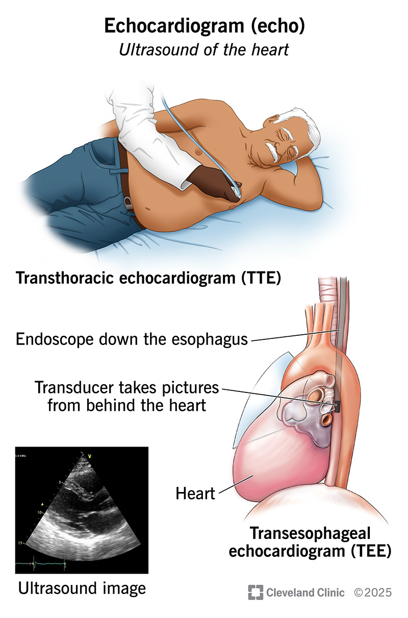
How is a transthoracic echocardiogram done?
A transthoracic echo is the type most people think of when they hear “heart echo.” It’s also the type most often used. It’s performed outside patient’s body.
A sonographer places a hand-held wand (called a transducer) on the outside of patient’s chest to send sound waves to the heart. These sound waves bounce off the different parts of the heart.
These “echoes” then appear as pictures on the sonographer’s computer screen. These pictures can also be saved for cardiologist and physician to review later.
Preparing for a transthoracic echo
There’s not much patient need to do to prepare for this type of echo. In general:
- Patient don’t need to avoid eating or drinking before a transthoracic echo.
- Patient can take his/her medications as usually do.
- Wear anything patient would like.
What to expect during a transthoracic echo
A transthoracic echo includes the following steps:
- Patient will be asked to remove clothing from the waist up and will be put on a hospital gown.
- Sonographer will place several electrodes on patient’s chest. These are small, flat, sticky patches. The electrodes are attached to an electrocardiograph (EKG) monitor. The EKG records patient’s heart’s electrical activity during the test.
- Patient will lie down on an exam table. Your sonographer will ask you to lie on your left side if possible.
- Sonographer will place a sound-wave transducer (wand) on several areas of Patient’s chest. There’s a small amount of gel on the end of the wand, which won’t harm the skin. This gel helps produce clearer pictures.
- Patient may hear swishing sounds; it means patient hearing blood flowing through his/her heart as the wand picks up the sound.
- Throughout the test, the sonographer may ask patient to hold his/her breath for several seconds at a time. Patient may also need to move into a different position.
Patient should feel no major discomfort during the test. Patient may feel a coolness on the skin from the gel on the wand. Patient may also feel a slight pressure of the wand against the chest.
How is a transesophageal echocardiogram done?
A transesophageal echo takes pictures from inside the chest, rather than from the outside. It can show Patient’s heart and valves in greater detail than a transthoracic echo. That’s because our body’s bones and tissues aren’t in between the transducer and our heart.
For this test, the sonographer guides a small transducer down the throat and esophagus using a long, flexible tube. This minimally invasive procedure may cause mild, temporary discomfort. But it has a low risk of serious problems.
This type of echo may be used:
- When a doctor needs a detailed look at aorta or the back of heart (especially left atrium or left ventricle).
- To check for blood clots.
- To evaluate patient’s mitral valve or aortic valve.
- If patient have obesity or lung disorders.
- If a transthoracic echo isn’t possible for any other reason(s).
Preparing for a transesophageal echo
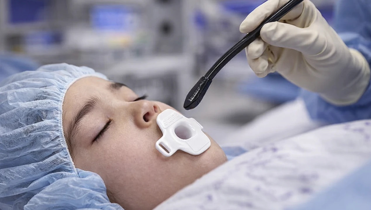 As patient prepare for your echo, he/she should tell the doctor if he/she has:
- Problems with esophagus, like a hiatal hernia etc.
- Problems swallowing.
- Sleep apnea, Sleep issues.
- IV drug use.
- Taking medication (especially allopathic).
- Anxiety.
- Pain.
What a doctor should say to the patient:
Preparations for the day of your test include:
- Find someone to drive you home from your test. This is because you’ll be sedated for the test, and you won’t be able to drive for 24 hours.
- Don’t eat or drink anything for at least six hours before your test. Your doctor may give you more specific instructions for eating and drinking. It’s essential you follow these closely.
- Ask your doctor about when to take your usual medications. You may be able to take them at your usual time with a small sip of water.
- Ask your doctor about when and how to take your diabetes medication, if this is relevant to you.
- Plan to leave any valuable personal items at home. You’ll have access to a storage locker for your belongings during the test.
- Plan to wear whatever’s most comfortable for you. You’ll change into a hospital gown before the echo begins.
What to expect during a transesophageal echo
A transesophageal echo includes the following steps:
- Patient will remove your clothing from the waist up and put on a hospital gown.
- Technician will place electrodes on patient’s chest. The electrodes allow the sonographer to monitor patient’s heart’s electrical activity during the test.
- Technician will place a blood pressure cuff on patient’s arm and a pulse oximeter on patient’s finger.
- Patient’s will gargle with a solution that numbs his throat. Technician will also spray patient’s throat with pain-relieving medication.
- To make patient more comfortable, patient be hooked up to an IV and sedated. Patient soon start to feel sleepy.
- Patient may have a tube in his/her nose that provides oxygen.
- Patient will lie on his left side on the exam table.
- The technician will insert an endoscope into patient’s mouth. This is a long, thin, flexible tube that has a transducer on the tip. The tube travels into patient’s throat and esophagus. It’s lubricated to help it slide down more easily. While this may feel uncomfortable, it won’t harm. Patient may need to swallow to move the transducer into the right spot (just behind the heart).
- Technician will take pictures. Patient’ won’t feel anything while this happens.
- After the technician has the pictures they need, they’ll remove the tube from patient’s throat. Patient will soon be able to get dressed and get ready to leave.
How is an exercise stress echocardiogram done?
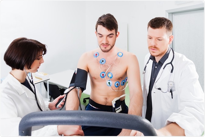 An exercise stress echo, sometimes simply called a stress echo, shows how patient’s heart works when it’s taxed. The test resembles a traditional exercise stress test. A technician will monitor patient’s heart rate and rhythm as well as patient’s blood pressure (this is standard during a stress test). But doctor will also use echo imaging (which isn’t normally used during a stress test).
This test shows how well patient’s heart can withstand activity. The sonographer takes pictures before patient start exercising and then right after he/she is done.
In some cases, patient won’t exercise. Instead, technician will give patient medication to make his/her heart work harder as if he/she were exercising. The goal is to force patient’s heart to need more oxygen.
When patient’s heart is under stress, sonographer can see details they might not be able to see if patient were lying on the exam table. These include problems with patient’s coronary arteries or the lining of patient’s heart.
Preparing for an exercise stress echo
Sonographer will give detailed instructions to the patient on how to prepare for the test. An exercise stress echo needs more preparation than other types of echo testing. These include:
- Not eating or drinking anything except water for at least four hours before the test.
- Not smoking on the day of test.
- Avoiding caffeine for 24 hours before the test. This includes caffeine in any form (coffee, tea, decaf drinks, sniffing, chewing tobacco and some over-the-counter pain medicines).
Ask your patient when and how to take his usual medications. Patient may need to avoid taking certain allopathic heart medications on the day of test. Patient may also need to change the dose of diabetes medication (allopathic).
Patient won’t be sedated, but he/she still may want to ask someone to drive him/her to and from the appointment. Patient may feel tired after the test.
Plan to wear comfortable clothes and shoes. Patient will need to walk or ride a stationary bike during the test, so wear what feels good.
What to expect during an exercise stress echo
The exercise stress echo will include the following steps:
- Sonographer will place electrodes on patient’s chest. These stickers are hooked up to an EKG monitor to check patient’s heart rate and rhythm during the test.
- Technician will measure patient’s heart rate, heart rhythm and blood pressure before he/she starts moving.
- Patient will lie on an exam table so the sonographer can take pictures of his/her heart. They’ll place a hand-held wand (the kind usually used for echo tests) on the outside of patient’s chest in various spots.
- Then, it’s time to start moving. Patient will walk on a treadmill or pedal on a stationary bike. The intensity will gradually increase. Patient will keep going until he/she is exhausted. This usually takes seven to 12 minutes.
- As patient is exercising, a technician will ask how he/she is feeling. Sonographer will also watch patient’s heart on the EKG monitor.
- If patient will stop moving, and he/she will have another echo test done.
- Patient will then do a short cool-down (slow walking or cycling), and the provider will monitor patient’s vitals until they’re back to normal.
If patient were given medication to stress his/her heart, the process will be a bit different. Patient won’t be on a treadmill or bike.
Results and Follow-Up
How do I get the results of my test?
After your cardiologist reviews your test, they’ll enter the results into your electronic medical record. Your primary care provider will have access to the results, too. You’ll discuss the results with one or both of these providers.
Ask any questions you’d like about the pictures and what they mean. Your provider will explain what the pictures show and whether you need follow-up tests or treatment.
Additional Common Questions
What should a I (patient) not do before an echocardiogram?
It depends on which type of echo you’re having done. Check with your provider to learn exactly what you should avoid. Things you may need to avoid before your echo include:
- Eating or drinking.
- Smoking or using any nicotine products.
- Drinking coffee or anything with caffeine in it. This includes colas, decaf drinks, which still contain a small amount of caffeine. It also includes over-the-counter medications that contain caffeine.
You may need to adjust your medication schedule before your echo. Don’t stop taking any medications or make any changes until you talk with your doctor.
P. S: This article is only for doctors and students having good knowledge about Homeopathy and allopathy.

For proper consultation and treatment, please visit our clinic.
 Dr. Sayyad Qaisar Ahmed (MD {Ukraine}, DHMS), Abdominal Surgeries, Oncological surgeries, Gastroenterologist, Specialist Homeopathic Medicines.
Senior research officer at Dnepropetrovsk state medical academy Ukraine.
Location: Al-Haytham clinic, Umer Farooq Chowk Risalpur Sadder (0923631023, 03119884588), K.P.K, Pakistan.
Find more about Dr Sayed Qaisar Ahmed at:
https://www.youtube.com/Dr Qaisar Ahmed