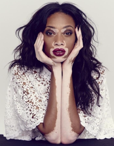
Vitiligo is a type of skin condition that occurs when the melanocytes in the body are destroyed. Melanocytes are the cells that are responsible for producing a pigment known as melanin, which gives the skin its color. When melanocytes fail to produce melanin, the area affected begins to turn white and lose its color. The areas that are affected by the condition and lose their pigment are known as macules.
Vitiligo occurs when the body’s immune system destroys the melanocyte cells. Apart from skin, this condition can also affect the hair and eyes. Any part of the body that has hair and is affected by this condition may turn silver or white.
The main pigment responsible for the hair and skin color is melanin. Melanocytes are the cells involved in the formation of melanin. Vitiligo is a dermatological condition (depigmentation) characterized by white spots or patches that appear in the body.
Lack or absence of melanin is often responsible for the appearance of the white patches. Improper functioning or destruction of the melanocytes may interfere with the cell’s ability to produce melanin. The condition can also affect your hair, scalp, eyebrows, and eyelashes, resulting in premature graying or whitening.
Vitiligo and Leukoderma Difference
Vitiligo can be localized (only one or very few body parts are affected), generalized (affecting many body parts) or segmented (the condition affects only one side of the body). An autoimmune condition such as Diabetes (Type-1) or Hashimoto’s thyroiditis may trigger the destruction of the melanocytes. Vitiligo may also be a hereditary condition. In some cases, stress or exposure to harmful chemicals may be responsible for the destruction of the melanocytes.
While many people confuse vitiligo with leukoderma, there is a small difference between the two. Like vitiligo, the white watches in leukoderma appear due to the destruction of the melanocytes. However, the destruction of the melanocytes in the case of leukoderma may result from an accident, a cut, burn or tear.
Vitiligo Causes
The exact causes of vitiligo remain unknown. However, various studies are being conducted to find what causes the lack of melanin in the body. Various researchers believe that this condition is an autoimmune disorder that occurs when the body starts attacking its cells. As per studies, some of the possible risk factors leading to the development of vitiligo may include the following,
Genetic factors
Certain studies have shown that around 30% of cases of vitiligo are genetic. This indicates that vitiligo condition is a hereditary problem and one can develop it if they have a family history. 
Genetic changes
Another potential risk factor of vitiligo is genetic changes in the DNA or genetic mutations, which can also affect the way melanocytes function. There are more than 30 genes in the body that may increase a person’s risk of developing vital conditions.
Autoimmune condition
Autoimmune condition is when the immune system of the body mistakes the body cells to be foreign invaders, such as microorganisms, and starts attacking the body’s cells. As a result, the immune system overreacts and produces antibodies to destroy its cells.
Researchers believe that it is a type of autoimmune disorder. Various autoimmune diseases may be linked to the development of Vitiligo. These autoimmune disorders may include the following:
- Rheumatoid arthritis,
- Type 1 diabetes,
- Alopecia areata,
- Thyroiditis,
- Lupus,
- Scleroderma,
- Addison’s disease,
- Psoriasis,
- Pernicious anemia.
Environmental factors
The various environmental factors that may increase the risk of developing vitiligo. These environmental factors include skin trauma, sunburns, emotional distress, exposure to UV radiation, and exposure to chemicals.
Oxidative stress
Oxidative stress due to an imbalance of antioxidants and oxygen molecules may also lead to an increased risk of developing vitiligo in some individuals.
Types of Vitiligo:
The various types of vitiligo. The location of the vitiligo, along with the prevalence of its symptoms, may differ depending on the type of vitiligo. The various types of vitiligo include the following,
1) Non-Segmental Vitiligo
Non-segmental vitiligo is one of the most commonly occurring types of Vitiligo, in which the patches are visible on both sides of the body and are usually symmetrical. The spots and patches are commonly found to have developed in body areas exposed to trauma, pressure, or sunburns. The five subcategories of non-segmental vitiligo include the following: 
- Generalized: In this type, macules appear on various areas of the body. This is also the most common type of vitiligo.
- Mucosal: In this type of vitiligo, the mucous membranes present in the mouth and the genital are affected by the condition.
- Focal: This type of vitiligo is seen most commonly in children. In this type, the patches form on smaller areas of the body.
- Acrofacial: In this type, the patches affect the face, fingers, and toes.
- Universal: The universal type of Vitiligo rarely occurs in which discolored patches cover most of the body areas.
2) Segmental Vitiligo
The segmental type of Vitiligo typically affects only one side of the body, such as the face or the hands. This type of vitiligo stops growing after the initial patch has been formed on the skin.
3) Mixed Vitiligo
In this type of vitiligo, the individual experiences a combination of both non-segmental and segmental vitiligo types.
4) Hypochromic Vitiligo
Hypochromic Vitiligo, also known as vitiligo minor, is a type characterized by the presence of a few white patches spread across the scalp and trunk. This particular type of vitiligo is commonly seen in people having darker skin tones.
Complications of Vitiligo
While vitiligo is more of a cosmetic concern, it can lead to various complications if not treated properly. The various complications of vitiligo include the following:
Sunburns:
As vitiligo condition causes a lack of melanin in the body, the skin becomes vulnerable to the harmful effects of the sun. Hence, individuals with wide go are more prone to getting sunburns.
Predisposition to autoimmune conditions:
Individuals are at a risk of developing other autoimmune conditions. Some of these autoimmune conditions may include anemia, diabetes and hypothyroidism.
Eye problems:
Individuals having vitiligo may experience eye issues such as inflammation of the iris or uveitis (a condition characterized by inflammation of the middle layer of the eye). Additionally, the patient may also experience a change in the color of the iris and may also have retina abnormality. 
Ear problems:
In certain cases, individuals with Vitiligo may experience hyperacusis (partial loss of hearing).
Psychological issues:
One of the most common complications of Vitiligo is issues with the way the patient’s skin looks. Individuals having Vitiligo may often experience low self-esteem and embarrassment due to their skin condition. This can also cause the patient to feel anxious and depressed. In some cases, the patient may also isolate themselves.
How to Diagnose Vitiligo?
The primary step in diagnosing vitiligo is a physical examination. Upon suspecting Vitiligo, check the symptoms on the body. During physical examination, conduct a medical history examination and check and ask your patient for any autoimmune and or any other skin infections, any minor/major injuries or accident that patient may have/had few weeks earlier. It is essential to ask your patient about any factor that may have led to the development of such diseases, skin trauma, contact with dirty water like sanitation or wastes from factories, sunburn etc.
It Is also important to ask your patient about family history of Vitiligo or other skin diseases, if any.
Apart from a physical and medical history examination, a doctor may also conduct another test using an ultraviolet lamp. The ultraviolet lamp (Wood’s lamp) helps the doctor look at the vitiligo patches and establish the difference between them and other skin conditions.
In some cases, also order a few blood tests and a skin biopsy. A skin biopsy will show if the pigment-producing cells (melanin) are present in the body areas affected by vitiligo.
Differences
Pityriasis Alba vs Vitiligo
Pityriasis Alba and Vitiligo may seem similar, but they are too distinct skin conditions. Although they have certain similarities in their appearances. The differences between Pityriasis alba and Vitiligo are given in the table below:
| S. No | Pityriasis Alba | Vitiligo |
| 1 | Pityriasis Alba is not a type of autoimmune condition. | Vitiligo is an autoimmune condition. |
| 2 | The changes in the skin occurring due to Pityriasis alba have faded borders. | The changes in the skin occurring due to vitiligo have extremely sharp borders. |
| 3 | The skin color of individuals having Pityriasis alba transition gradually | The changes in skin color in the case of vitiligo happen abruptly. |
Leucoderma vs Vitiligo
Both leucoderma and Vitiligo are two commonly occurring skin conditions that are characterized by the appearance of white spots on the skin. In many instances, it is difficult to identify whether the condition affecting is Vitiligo or leucoderma. While the condition shares certain similarities, they also have certain differences setting them apart. The differences between leukoderma and Vitiligo are given below:
| S. No | Leucoderma | Vitiligo |
| 1 | Leucoderma is not an autoimmune condition and can be caused due to various problems such as chemical exposure, trauma, infections, burns, injuries, allergic reactions, etc. | Vitiligo is an autoimmune condition and its exact causes of vitiligo are not known. However, a combination of autoimmune, genetic factors and environmental factors may act as its risk factors. |
| 2 | Various pigment disorders that come under leucoderma, such as Chemical Leucoderma, post-inflammatory hypopigmentation, piebaldism, halo nevus, nevus depigmentosus, etc. | Vitiligo has two major types: segmental vitiligo and non-segmental vitiligo. |
| 3 | The progression of Leucoderma is based on the underlying cause. In case the underlying reason for Leucoderma is progressive, the depigmentation process in the body may continue. | Vitiligo is typically progressive and can spread over time. However, the extent and rate of progression may vary from patient to patient. While vitiligo may progress quickly in some cases, it may be slow in others. |
| 4 | The treatment for leucoderma is given depending on its cause, the stage, the patient’s overall health and age. | The aim of treatment in the case of vitiligo is to restore the pigment in the areas affected or even the skin pigment out. |
My experience
In my (Dr Qaisar Ahmed) opinion, it is some kind of a viral skin infection; in my (Dr Qaisar Ahmed) 27 years of practice I had seen hundreds of vitiligo patients who had the disease after some type of injuries especially road, dirty water, sanitation water etc.
Albinism Vs Vitiligo
Both albinism and vitiligo are two conditions that affect the pigment on the skin. Although both conditions appear similar, leading to confusion between them, there are several differences between the two conditions. The differences between albinism and vitiligo are listed below,
| S. No | Albinism | Vitiligo |
| 1 | Albinism is a genetic disorder. | Vitiligo is an autoimmune condition. |
| 2. | Individuals with albinism have very little pigment or no pigment at all on their skin at the time of birth. | Individuals having vitiligo experience white-colored patches on their bodies. These patches may develop on one or both the sides of the body and may also be symmetrical. These patches can develop in any area of the body. |
| 3 | Albinism is a condition that is inherited and occurs as a result of mutation in one of the genes responsible for the production of melanin in the body. When the proteins responsible for melanin production are not available in the body or are deformed, melanin cannot be produced. This eventually leads to the loss of colour of the skin. | Vitiligo is caused when the activity of the immune system destroys the body’s cells responsible for producing melanin, called melanocytes. This leads to the formation of white patches on the body. |
Hypopigmentation Vs Vitiligo
Hypopigmentation and vitiligo are two skin conditions that are characterized by the formation of skin patches that are lighter in color when compared to the rest of the body. Although Vitiligo and hypopigmentation have similar symptoms, they are two distinct conditions. The various differences between hypopigmentation and vitiligo are listed in the table below.
| S. No | Hypopigmentation | Vitiligo |
| 1 | Hypopigmentation is a general term that is used to define reduced skin pigmentation or color. | Vitiligo is a lifelong condition. |
| 2 | Hypopigmentation can develop on its own or occur due to various causes such as infections, skin disorders, certain health conditions, injuries and effects of certain medications. Hypopigmentation may also be present at birth or may also develop later on in life. | Vitiligo is an autoimmune condition in which the immune system attacks and destroys the melanocytes in the body, which are responsible for the production of a pigment called melanin. As a result of Vitiligo, White colored patches develop on the skin. |
| 3 | The treatment for hyperpigmentation depends upon various factors such as the patient’s age, the area of the skin affected, the overall health of the patient and the underlying cause of the problem. Additionally, in certain cases where hypopigmentation occurs as a result of an injury, cut or burn, the hypopigmentation may resolve on its own as the skin heals. | There is no cure available for Vitiligo. The treatment given for vitiligo aims to add pigment or color to the depigmented patches on the skin, as well as slow down the progression of the condition. |
| 4 | The types of hypopigmentation include: Generalized and localized. | The two major types of vitiligo include: Segmental vitiligo and non-segmental vitiligo. |
Nevus Depigmentosus vs Vitiligo
Both Nevus Depigmentosus and vitiligo are conditions involving depigmentation of the skin. While the conditions look similar, they have distinct characteristics that set them apart. The various differences between Nevus Depigmentosus and vitiligo include the following,
| Nevus Depigmentosus | Vitiligo | |
| 1 | Nevus Depigmentosus is a type of congenital condition that is present in both. | Vitiligo is a type of autoimmune condition characterized by the presence of depigmentic patches on the skin. |
| 2 | The patches occurring in Nevus Depigmentosus are usually localized to one particular area. | The depigmented patch in case of vitiligo may occur anywhere on the body. It can affect either one or both sides of the body. |
| 3 | The patches in Nevus Depigmentosus don not spread or change its size and shape significantly over time. | The patches in case of vitilgo may spread or enlarge with time. |
Leprosy Vs Vitiligo
Leprosy and vitiligo are two types of conditions affecting the skin. Both vitiligo and leprosy are distinct skin conditions. The various differences between these two conditions are listed below,
| S. No | Leprosy | Vitiligo |
| 1 | Leprosy is a type of infectious condition that occurs due to bacteria. | Vitiligo is a type of autoimmune condition that leads to the loss of the pigment called melanin from the skin. |
The symptoms of leprosy include the following,
|
The symptoms of vitiligo include the following,
|
|
| 3 | Leprosy is contagious and can be transferred from one individual to another individual through the respiratory droplets released in the air by the infected person. |
Vitiligo allopathic treatment
There are various treatments available to treat vitiligo. The treatment given for vitiligo may vary depending on the effect of the condition on the body and the body’s response to the treatment. In certain cases, the treatment goal may be to add pigment to the body or restore the color balance in the skin, while some treatments may aim to remove the pigment. 
To treat vitiligo, medicines, surgery, or a combination of both approaches may be prescribed. In general, the treatment for vitiligo may include:
- Medicines.
- Topical creams.
- Ultraviolet A (PUVA) light therapy.
- Depigmentation.
- Narrowband ultraviolet B light therapy.
- Excimer laser treatment.
- Surgery (skin grafts and blister grafting).
Homeopathic Treatment for Vitiligo & Leukoderma
When it comes to treatment, many people prefer Homeopathy over conventional treatments and medications. Being natural, curable and with no side effects are the main causes of Homeopathic treatment. People with vitiligo and leukoderma have benefitted immensely from the treatment.
Homeopathic medicines activate the melanocytes. This activation triggers the formation of the melanin, thereby resulting in re-pigmentation.
Here are few best proven Homeopathic medicines for these diseases:
Sulphur
Dry, scaly, unhealthy; every little injury suppurates. Freckles. Tinea versicolor or pityriasis versicolor. Itching, burning; worse scratching and washing. Pimply eruption, pustules, rhagades, hangnails. Excoriation, especially in folds. Feeling of a band around bones. Skin affections after local medication. Pruritus, especially from warmth, is evening, often recurs in springtime, in damp weather.

Zincum Metalicum
Formication of feet and legs as from bugs crawling over the skin, preventing sleep. Eczema, especially in the anemic and neurotic. Itching of thighs and hollow of knees. Retrocession of eruptions. Tinea versicolor or pityriasis versicolor.
Sepia Officianalis
Herpes circinatus in isolated spots. Itching; not relieved by scratching; worse in bends of elbows and knees. Chloasma; herpetic eruption on lips, about mouth and nose. Tinea versicolor or pityriasis versicolor. Ringworm-like eruption every spring. Urticaria on going in open air; better in warm room. Hyperhidrosis and bromhidrosis. Sweat on feet, worse on toes, intolerable odor. Lentigo in young women. Ichthyosis with offensive odor of skin.
Mezereum
Eczema; intolerable itching; chilliness with pruritus; worse in bed. Tinea versicolor or pityriasis versicolor. Ulcers itch and burn, surrounded by vesicles and shining, fiery-red areola. Zona, with burning pain. Bones, especially long bones, inflamed and swollen; caries, exostosis; pain worse night, touch, damp weather. Eruptions ulcerate and form thick scabs under purulent matter exudes (Chrysophanic acid).
Thuja Occidentalis
Polypi, tubercles, warts epithelioma, naevi, carbuncles; ulcers, especially in ano-genital region. Freckles and blotches. Perspiration sweetish, and strong. Dry skin, with brown spots. Zona; herpetic eruptions. Tinea versicolor or pityriasis versicolor. Glandular enlargement. Nails crippled; brittle and soft. Eruptions only on covered parts; worse after scratching. Very sensitive to touch. Coldness of one side. Sarcoma; polypi. Brown spots on hands and arms.
Mercuris Solubus
Almost constantly moist. Persistent dryness of the skin contra indicates mercurius. Excessive odorous viscid perspiration; worse, night. General tendency to free perspiration, but patient is not relieved thereby. Vesicular and pustular eruptions. Tinea versicolor or pityriasis versicolor. Ulcers, irregular in shape, edges undefined. Pimples around the main eruption. Itching, worse from warmth of bed. Crusta lactea; yellowish-brown crusts, considerable suppuration. Glands swell every time patient takes cold. Buboes. Orchitis.
Clematis Erecta
Red, burning, vesicular, scaly, scabby. Itches terribly; worse, washing in cold water; worse face and hands and scalp around occiput. Glands hot, painful, swollen; worse inguinal glands. Glandular indurations and tumors of breast. Varicose ulcers. Tinea versicolor or pityriasis versicolor.
Tellurium Metallicum
Itching of hands and feet. Herpetic spots; ringworm. Ring-shaped lesions, offensive odors from affected parts. Barber’s itch. Stinging in skin. Fetid exhalations. Offensive foot-sweat. Eczema, back of ears and occiput. Circular patches of eczema. Tinea versicolor or pityriasis versicolor.
Thyroidinum
Psoriasis associated with adiposity (not in developing stage). Skin dry, impoverished. Cold hands and feet. Eczema. Uterine fibroids. Browne swelling. Swelling of glands of stony hardness. Sluggish cases. Jaundice with pruritus. Ichthyosis, lupus. Itching without eruption, worse night.
Kalium Sulphuricum
Psoriasis. Eczema; burning, itching, papular eruption. Nettle-rash. Polypi. Epithelioma. Seborrhoea. Favus. Ringworm of scalp or beard with abundant scales.
Hamamelis Virginiana
Bluish chilblains. Phlebitis. Purpura. Varicose veins and ulcers; very sore. Burns. Ecchymosis. Traumatic inflammations.
Calceria Carbonicum
Unhealthy; readily ulcerating; flaccid. Small wounds do not heal readily. Glands swollen. Nettle rash; better in cold air. Warts on face and hands. Petechial eruptions. Chilblains. Boils.
Arsenicum Album 
Itching, burning, swellings; oedema, eruption, papular, dry, rough, scaly; worse cold and scratching. Malignant pustules. Ulcers with offensive discharge. Desquamation of the skin of the body. Anthrax. Poisoned wounds. Urticaria, with burning and restlessness. Psoriasis. Scirrhous. Icy coldness of body. Epithelioma of the skin. Gangrenous inflammations.
Arsenic Sulphuratum
Leucoderma and squamous syphilides.
Berberis Aquifolium
The top recommendation for clearing acne scars, reducing dark spots, and bringing a natural glow. Berberis Aquifolium can be used in all the cases where the skin is dark or has scars due to acne or any other conditions. This medicine never fails to bring a glow to the skin and lighten the complexion. I consider it as the first choice of medicine in cases where a clear facial complexion is desired. It has shown very promising results and given complete satisfaction to my patients as well as me. To sum up, I would call Berberis Aquifolium a ‘precious gem’ in homeopathy to attain fair, clear and glowing skin.
Antimuonium Crudum
Itching. Eruptions, measles and/or chickenpox-like eruption, miliary eruptions and nettle-rash. Tumors and blisters. Thick, hard scabs, often honey-yellow, here and there a crack oozing a green sanious fluid, burning. Urticaria white, with red areolae, which itch fearfully. Pustules with yellowish or brown scurf. Freckles. Hepatic spots. Deep spongy ulcers with gastric ulcers. Fistulous ulcers. Horn-like excrescences and disposition to abnormal organizations of the skin. Corns and callous excrescences on the feet. Nails discolored and deformed. Red and hot swellings. Degeneration of the skin. Fungus of the joints.
Antimuonium Tartaric
Itching in the skin. Itching pimples, and miliary eruption. Eruptions like scabies. Eruption of pustules, like varioloids, filled with pus, with red areola (like smallpox), and which afterwards form a crust, and leave a white scar. Itching round inveterate ulcers. Pustular eruption on different portions of the body, leaving a bluish-red mark.
Santalum Album
Used to improve skin complexion and reduce blemishes.
Medorrhinum
Yellow. Intense and incessant itching; worse night and when thinking of it. Fiery red rash about anus in babies. Copper-colored spots. Favus. Tumors and abnormal growth.
Psorinum
Dirty, dingy look. Dry, lusterless, rough hair. Intolerable itching. Herpetic eruptions, especially on scalp and bends of joints with itching; worse, from warmth of bed. Enlarged glands. Sebaceous glands secrete excessively, oily skin. Indolent ulcers, slow to heal. Eczema behind ears. Crusty eruptions all over. Urticaria after every exertion. Pustules near fingernails.
Psorinum has shown the best results where itching is intolerable, and the skin gets raw or may even start to bleed from intense and continuous scratching.
Acid Nitricum
Dryness of the skin. Itching urticaria. Blackness of pores. Brown sphacelus. Reddish-brown spots (scattered over body, esp. if in dark-haired people) and deep-colored ephelis on skin. Copper or violet-colored spots. Itching tetters. Pimples, or exanthema in general; stinging exanthema. Pain from chilblains and corns on feet. In a moderately cold temperature limbs become as if frozen, inflamed, and itching, and skin cracks. Large furunculi. Mercurial ulcers.
Silicia Tera
Felons, abscesses, boils, old fistulous ulcers. Delicate, pale, waxy. Cracks at end of fingers. Painless swelling of glands. Rose-colored blotches. Scars suddenly become painful. Pus offensive. Promotes expulsion of foreign bodies from tissues. Every little injury suppurates. Long lasting suppuration and fistulous tracts. Dry finger tips. Eruptions itch only in daytime and evening. Crippled nails. Indurated tumors. Abscesses of joints. After impure vaccination. Bursa. Lepra, nodes, and coppery spots. Keloid growths.
Staphisagria
Tingling, as from insects, over whole body. Chronic miliary eruptions, sometimes with convulsive jerks. Eczema; thick scabs, dry, and itch violently; scratching changes location of itching. Herpetic eruptions, with itching in evening; and burning sensation after scratching them. Tinea Versicolor. Arthritic nodosities on the joints. Dry, crusty tetters on the joints. Painful engorgement and induration of the glands. Unhealthy skin, easily suppurating. Frequent furunculi. Ulcers, with tearing shootings (gnawing pains), or itching smarting. Jerking and tearing round ulcers. Wens and encysted tumors burst after Staphisagria.
Natrum Muriaticum
Greasy/oily skin and hairs. Dry eruptions. Itching and pricking in skin. Rash over whole body, with stinging sensation in skin. Pityriasis. Fever blisters. Urticaria; itch and burn. Crusty eruptions in bends of limbs, margin of scalp, behind ears. Cracks in the skin. Warts on palms of hands. Eczema; raw, red, and inflamed; worse, eating salt, at seashore. Affects hair follicles. Alopecia. Hives, itching after exertion. Herpes circinate. Leucocythemia/leukemia. Seborrhea.
Phosphorus
Ecchymosis (purple large flat patch on skin). Ephelis (brown and or white patches on skin). Odour of body, changed. Fungus haematodes (an archaic 19th-century medical term for a highly vascular, soft malignant tumor, most commonly identified as retinoblastoma or similar encephaloid (brain-like) cancers. It was historically described as “soft cancer” or a bleeding fungus-like growth, often appearing in the eye or extremities). Wounds bleed very much, even if small; they heal and break out again. Jaundice. Little ulcer outside of large ones. Petechiae. Ecchymosis. Purpura hemorrhagic. Scurvy. 
Borax Veneta
Psoriasis. Erysipelas in face. Itching on back of finger-joints. Herpes. Erysipelatous inflammation with swelling and tension. Chilblains relieved in open air. Trade eruptions on fingers and hands, itching and stinging. Ends of hair become tangled/Plica-polonica. Psoriasis. Skin difficult to heal; dingy, unhealthy, every injury tends to ulceration. Erysipelatous inflammations, with swelling and tension of the part affected. Tinea Versicolor or Pityriasis Versicolor. Erysipelatous inflammation, with chilliness, followed by heaviness and pulsation in the head; later, bleeding of the nose. Tendency of old wounds to suppurate. Whitish pimples, with red areola. Purulent and phagedenic vesicles.
Baryta Carbonicum
Arterial fibrosis. Blood-vessels soften and degenerate, become distended, and aneurisms, ruptures, and apoplexies result. degenerative changes. Sensation in different parts, like the pricks of burning needles, itching and crawling sensations. Intolerable itching and tingling over the whole body at night. Excoriation and oozing in several parts of the skin. Injuries in the skin healing with difficulty. Swelling and induration of the glands. Whitlows.
Baryta Muriaticum
Pricking in the skin. Burning and pricking in excoriated places. Small itchy eruptions. Glands inflamed and ulcerated. Hemorrhages. Tinea Versicolor or Pityriasis Versicolor.
Kalium Bromatum
Nodular form of chronic gout. Acne of face, pustules. Itching; worse on chest, shoulders, and face. Anaesthesia of skin. Psoriasis.
Mercurius Solubilis
Almost constantly moist. Excessive odorous viscid perspiration; worse, night. General tendency to free perspiration, but patient is not relieved thereby. Vesicular and pustular eruptions. Ulcers, irregular in shape, edges undefined. Pimples around the main eruption. Itching, worse from warmth of bed. Crusta lactea; yellowish-brown crusts, considerable suppuration. Glands swell every time patient takes cold. Buboes. Orchitis.
Kalium Bromatum
Acne of face, pustules. Itching; worse on chest, shoulders, and face. Anesthesia of skin. Psoriasis. Sebaceous cysts. Seborrhea.
Lycopodium Clavatum
Skin ulcers. Abscesses beneath skin. Hives. Violent itching; fissured eruptions. Acne. Chronic eczema associated with urinary, gastric and hepatic disorders, bleeds easily. Skin becomes thick and indurated. Varicose veins, naevi, erectile tumors. Brown spots, freckles worse on left side of face and nose. Dry, shrunken, especially palms; hair becomes prematurely gray. Dropsies. Offensive secretions; viscid and offensive perspiration. Psoriasis.
Psoralea Corylifolia (Babchi)
Sometimes used as a “mother tincture” for external application to stimulate re-pigmentation when exposed to sunlight.
Psorinum
Psorinum is the appropriate remedy for people with oily or greasy facial skin which has a dark and dirty look. The face may also show pimples due to highly active oil-producing sebaceous glands on the face. Here Psorinum will help to clear the pimples and dark skin and control the excessive oiliness of the skin. Psorinum will deep-cleanse the skin pores on the face making it oil-free, clear and glowing. High sensitivity towards cold air may be found in persons needing Psorinum. Offensive perspiration may also be a complaint. 
Psoralea Corylifolia (Babchi)
Sometimes used as a “mother tincture” for external application to stimulate re-pigmentation when exposed to sunlight.
Bovista
Facial pimples, dark skin or any other skin affection caused by the injudicious use of cosmetic products can benefit from the use of the remedy Bovista which is used to clear the skin. Bovista holds a significant place among homeopathy medicines for the treatment of skin problems consequent to the excessive use of cosmetics. Bovista helps in clearing facial skin and makes it look bright and glowing. It also removes pimples or other afflictions resulting from the use of cosmetics.
P. S: This article is only for doctors and students having good knowledge about Homeopathy and allopathy.

For proper consultation and treatment, please visit our clinic.
 Dr. Sayyad Qaisar Ahmed (MD {Ukraine}, DHMS), Abdominal Surgeries, Oncological surgeries, Gastroenterologist, Specialist Homeopathic Medicines.
Senior research officer at Dnepropetrovsk state medical academy Ukraine.
Location: Al-Haytham clinic, Umer Farooq Chowk Risalpur Sadder (0923631023, 03119884588), K.P.K, Pakistan.
Find more about Dr Sayed Qaisar Ahmed at:
https://www.youtube.com/Dr Qaisar Ahmed
https://www.facebook.com/ahmed drqaisar